Abdominal Birth Defects in Grand Rapids, Michigan
Abdominal birth defects are congenital (present at birth) conditions that affect a baby’s abdominal wall, diaphragm, and the position of internal organs. In Grand Rapids and throughout West Michigan, these conditions are rare but serious and require rapid, highly specialized care.
Families in the Grand Rapids area are often cared for at regional centers with advanced neonatal and pediatric surgery services, including:
- Spectrum Health Helen DeVos Children’s Hospital (Corewell Health)
- Trinity Health Grand Rapids
- University of Michigan Health–West (Metro Health)
- Mercy Health and affiliated regional hospitals
Because these defects can affect breathing and circulation within minutes of birth, delivery is usually planned at, or followed by transfer to, a hospital with a Level III or IV Neonatal Intensive Care Unit (NICU).
The three main abdominal birth defects are:
- Congenital diaphragmatic hernia (CDH) – abdominal organs protrude into the chest cavity
- Exomphalos (omphalocele) – organs protrude through the navel (umbilicus) in a protective sac
- Gastroschisis – organs protrude through an opening in the abdominal wall, usually next to the navel, without a protective sac
These conditions are usually detected during pregnancy and managed by a high‑risk obstetrics and neonatal team. In Kent County and the greater Grand Rapids region, pregnant patients are commonly referred to maternal–fetal medicine specialists for detailed diagnosis, counseling, and delivery planning.
How Abdominal Birth Defects Are Detected in Pregnancy
Prenatal Ultrasound and Screening in Grand Rapids
Most abdominal birth defects are first suspected during routine prenatal ultrasound. In the Grand Rapids area, these scans are typically performed in OB/GYN practices, community clinics, and hospital-based maternal–fetal medicine programs affiliated with:
- Spectrum Health / Corewell Health in Grand Rapids
- Trinity Health Grand Rapids
- University of Michigan Health–West (Metro Health)
- Mercy Health and other West Michigan partners
During an ultrasound, your provider may see:
- Abdominal organs appearing outside the baby’s body
- Abnormal positioning of the stomach, intestines, or liver
- A smaller or shifted lung area, suggesting a diaphragmatic hernia
- Signs of polyhydramnios – too much amniotic fluid around the baby
Because many families in West Michigan live in rural or lakeshore communities, early ultrasound at local clinics is often followed by referral into Grand Rapids for more detailed imaging and consultation.
Polyhydramnios as a Warning Sign
Polyhydramnios (too much amniotic fluid) can be a clue that something is affecting the baby’s ability to swallow or process fluid, including possible abdominal birth defects. When polyhydramnios is seen together with abnormal organ position, your provider may recommend:
- A detailed level II ultrasound
- Fetal echocardiogram to evaluate the baby’s heart
- Possible genetic testing
- Referral to a maternal–fetal medicine specialist in Grand Rapids
Families in Kent County can access high‑risk pregnancy services and counseling through major Grand Rapids health systems, and may receive additional support and referrals from the Kent County Health Department and Grand Rapids Public Health programs.
Causes and Prevention
Unknown Causes
For most cases of diaphragmatic hernia, exomphalos (omphalocele), and gastroschisis:
- The exact causes are unknown
- They are believed to result from abnormal development of the diaphragm or abdominal wall early in pregnancy
- There is no proven way to prevent these conditions
Researchers continue to study possible genetic and environmental factors, but most parents in Grand Rapids who have a baby with an abdominal birth defect:
- Have no known risk factors
- Did nothing to cause the condition
Public health messaging in Michigan emphasizes healthy pregnancy habits (prenatal vitamins, avoiding tobacco and substance use, regular prenatal care), which are important for overall fetal health but do not guarantee prevention of these specific defects.
Understanding the Diaphragm and Breathing
How the Diaphragm Works
The diaphragm is a thin, dome-shaped sheet of muscle that separates the chest cavity from the abdominal cavity. It:
- Contracts when we breathe in, pulling air into the lungs
- Relaxes when we breathe out, pushing air out of the lungs
Normally, all abdominal organs—such as the stomach, intestines, liver, and pancreas—remain below the diaphragm. If there is a hole or weakness in the diaphragm, organs can move into the chest and interfere with lung growth and breathing.
Congenital Diaphragmatic Hernia (CDH)
What Is a Diaphragmatic Hernia?
A congenital diaphragmatic hernia (CDH) occurs when there is an abnormal hole in the diaphragm. This opening allows abdominal organs to move into the chest cavity, where they can crowd the lungs and heart.
This crowding can:
- Limit lung growth and development before birth
- Make breathing very difficult immediately after delivery
- Lead to low oxygen levels in the baby’s body
Signs at Birth
Newborns with a diaphragmatic hernia may:
- Look blue or gray (cyanotic) because of low oxygen
- Breathe rapidly, erratically, or not at all
- Have a flatter-than-normal abdomen, because organs have moved into the chest
- Have decreased breath sounds on one side of the chest
In Grand Rapids, babies with suspected CDH are typically delivered at, or urgently transferred to, hospitals with Level III or IV NICUs, such as Spectrum Health Helen DeVos Children’s Hospital, where neonatal intensive care, pediatric surgery, and advanced breathing support (including ECMO in some cases) are available 24/7.
How Common Is Diaphragmatic Hernia?
In Michigan, congenital diaphragmatic hernia occurs in approximately 1 in every 2,500 births. This makes it a rare but serious condition that benefits from coordinated regional care across West Michigan.
Treatment for Diaphragmatic Hernia
CDH is a medical emergency at birth. Treatment usually includes:
- Immediate respiratory support (often a breathing machine/ventilator)
- Care in a specialized neonatal intensive care unit (NICU)
- Surgery to move the abdominal organs back into the abdomen and repair the hole in the diaphragm
Because Grand Rapids experiences long, cold winters and higher rates of respiratory illnesses such as RSV, influenza, and COVID‑19, care teams place strong emphasis on:
- Infection prevention in the NICU
- Vaccination of household contacts when appropriate
- Careful follow-up after discharge to reduce the risk of severe lung infections
Outlook and Long-Term Considerations
The outcome for a child with CDH depends largely on:
- How well the lungs developed before birth
- The size and location of the hernia
- Whether there are other health problems
Some children may:
- Be more prone to lung infections and breathing problems, especially during Michigan’s winter and early spring
- Need medications, inhalers, or supplemental oxygen
- Require follow-up care with pulmonologists, cardiologists, developmental specialists, and pediatric surgeons
Families in the Grand Rapids area can access long-term follow-up through local children’s hospitals, multidisciplinary clinics, and community programs that help coordinate appointments, therapies, and home care.
Exomphalos (Omphalocele)
What Is Exomphalos?
Exomphalos, also called omphalocele, is a birth defect where:
- There is an opening in the abdominal wall, usually at the navel
- The intestines and sometimes the liver and other organs protrude through this opening
- The protruding organs are covered by a thin protective sac made of the abdominal lining (peritoneum) and sometimes the amniotic membrane
Most defects occur at the umbilicus (belly button). In severe cases, the opening can be up to 10 cm in diameter, and organs may extend into the umbilical cord.
How Common Is Exomphalos?
In Michigan, exomphalos occurs in approximately 1 in every 2,500 births, similar to the rate of diaphragmatic hernia. Because Grand Rapids serves as a referral center for much of West and Northern Michigan, many families from outside Kent County may receive specialized care here.
Associated Conditions
Exomphalos is more likely than gastroschisis to be associated with other birth defects. It is estimated that:
- One-third to one-half of babies with exomphalos have additional problems, such as:
- Congenital heart defects
- Chromosomal abnormalities
- Other structural anomalies
Because of this, pregnant patients in Grand Rapids with a fetus diagnosed with exomphalos are often referred for:
- Fetal echocardiogram
- Genetic counseling and testing
- Consultations with neonatology and pediatric surgery teams
- Social work support to help plan for delivery, transportation, and possible extended NICU stays
Diagnosis
Exomphalos is usually diagnosed by:
- Prenatal ultrasound during pregnancy
- Physical examination at birth, as the defect is clearly visible
Local maternal–fetal medicine clinics in Grand Rapids may also use fetal MRI or additional imaging to better understand the size of the defect and plan for delivery.
Treatment for Exomphalos
After delivery in a hospital equipped with a NICU and pediatric surgeons:
- The protruding organs are carefully protected to prevent injury, drying, and infection.
- Surgery is typically performed within the first days of life to return organs to the abdomen and close the opening.
If there is not enough room in the baby’s abdomen to safely return all organs at once:
- A special protective sack (silo) is stitched around the organs.
- Over time, this sack shrinks, gently guiding the organs back into the abdomen.
- Once the organs are fully returned, the opening is sutured closed.
Hospital Stay and Follow-Up
Babies with exomphalos often require a prolonged hospital stay in a Grand Rapids NICU to:
- Monitor breathing and heart function
- Support feeding and nutrition (often starting with IV nutrition and then tube feeds)
- Watch for infection or digestive problems
Long-term follow-up may involve:
- Pediatric surgery for ongoing abdominal wall or scar issues
- Cardiology if heart defects are present
- Nutrition and feeding specialists, including occupational and speech therapists
- Early intervention programs to support growth and development
Families can access additional support through Kent County and Grand Rapids community resources, including home visiting programs, WIC, and early childhood services.
Gastroschisis
What Is Gastroschisis?
Gastroschisis is another type of abdominal wall defect where:
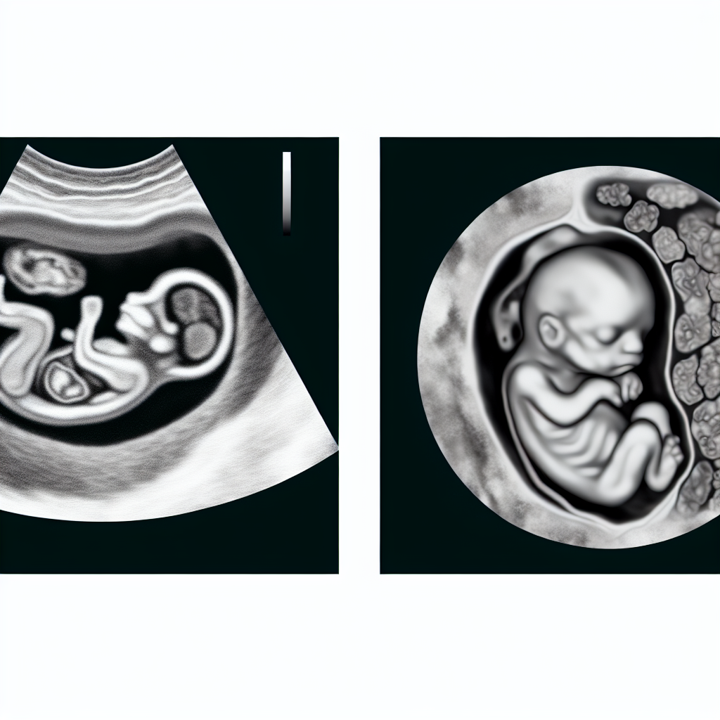 Normal Fetal Abdomen vs. Abdominal Birth Defect on Ultrasound
- The intestines and sometimes other organs protrude through an opening in the abdominal wall
- The opening is usually to the right of the navel, not through the navel itself
- Unlike exomphalos, the protruding organs are not covered by a protective sac—they are exposed directly to amniotic fluid and, after birth, the outside environment
How Common Is Gastroschisis?
In Michigan, gastroschisis occurs in approximately 1 in every 4,000 births. It is slightly less common than exomphalos and diaphragmatic hernia. Some studies have suggested higher rates in younger mothers, which is important for areas like Grand Rapids with diverse age and socioeconomic demographics.
Differences Between Gastroschisis and Exomphalos
Key differences include:
- Location:
- Gastroschisis – opening is next to the navel
- Exomphalos – opening is at the navel
- Covering:
- Gastroschisis – organs are not covered by a sac
- Exomphalos – organs are covered by a protective sac
- Associated defects:
- Gastroschisis – less likely to have other major organ problems
- Exomphalos – more often associated with heart and other defects
Risks and Immediate Care
Because the intestines are exposed in gastroschisis, they can:
- Dry out
- Become irritated or inflamed
- Be at higher risk for infection or injury
Immediately after birth in a Grand Rapids hospital:
- The protruding organs are covered with moist, sterile dressings or placed in a special sterile bag to protect them.
- The baby is stabilized in the NICU, and IV fluids, antibiotics, and careful temperature control are started.
In the most severe cases, almost all abdominal organs may be outside the body, requiring very careful planning and staged surgical repair.
Diagnosis
Like other abdominal birth defects, gastroschisis is usually diagnosed by:
- Prenatal ultrasound during pregnancy
- Physical examination at birth
Many families in West Michigan may first learn of the diagnosis at a community hospital and then be referred into Grand Rapids for ongoing prenatal care and delivery planning.
Treatment for Gastroschisis
Treatment depends on:
- How many organs are outside the abdomen
- Whether the intestines are damaged, twisted, or swollen
- How much space is available inside the baby’s abdomen
Two main approaches are used:
Primary closure
- If the baby’s abdomen is large enough, surgeons relocate the organs back inside the body and sew the opening closed shortly after birth.
Staged repair with a silo
- If the defect is large or the intestines are very swollen, a mesh or plastic sack (silo) is stitched around the organs.
- Over several days, the organs are gradually guided back into the abdomen.
- Once everything is in place, the abdominal wall is closed.
Possible Complications and Long-Term Care
If the intestines are damaged, the child may experience:
- Feeding difficulties
- Problems with digestion and nutrient absorption
- Need for long-term nutritional support, sometimes including IV nutrition (total parenteral nutrition, or TPN)
Given Michigan’s seasonal viral illnesses, especially in fall and winter, pediatric teams in Grand Rapids focus on:
- Careful infection prevention in the hospital and at home
- Regular follow-up with pediatric gastroenterology and surgery
- Support for families through hospital social work, home nursing when needed, and regional resources such as early intervention and nutrition programs
How These Defects Develop
During early fetal development, the abdominal wall and diaphragm must fully form and fuse. If this process is disrupted:
- A hole in the diaphragm can lead to congenital diaphragmatic hernia
- A defect at the navel can cause exomphalos (omphalocele)
- A defect next to the navel can cause gastroschisis
The organs of the digestive system—intestines, stomach, pancreas, liver—are all located in the abdomen and normally separated from the chest cavity by the diaphragm. When normal fusion and closure do not occur, these organs can protrude through the opening and may affect lung growth, circulation, and digestion.
Where to Get Help in Grand Rapids, Michigan
If an abdominal birth defect is suspected during pregnancy or after delivery, families in the Grand Rapids area have access to a strong regional network of maternal, newborn, and pediatric specialists.
You can seek help from:
- Your OB/GYN or family doctor
- Pediatrician or neonatologist
- Maternal–fetal medicine specialists at major Grand Rapids hospitals
- Pediatric surgeons at Spectrum Health Helen DeVos Children’s Hospital and other regional centers
Local and regional resources include:
- Kent County Health Department – information, referrals, immunization services, and public health support
- Grand Rapids Public Health and community health centers – assistance with prenatal care, follow-up appointments, and connections to specialists
- Hospital-based social workers and case managers – help with transportation, insurance questions, lodging (especially during winter travel), and emotional support for families coming from rural West Michigan or lakeshore communities
For many families, care involves coordination between local providers (in communities such as Holland, Muskegon, or Big Rapids) and specialists in Grand Rapids. Early communication with your care team helps ensure a safe delivery plan and timely transfer if needed.
Key Points About Abdominal Birth Defects in Grand Rapids
- Polyhydramnios (too much amniotic fluid) on prenatal ultrasound can be a sign of an abdominal birth defect and should prompt detailed evaluation.
- The exact causes are unknown, and there is currently no proven way to prevent diaphragmatic hernia, exomphalos, or gastroschisis. Parents did not cause these conditions.
- Newborns with diaphragmatic hernia may look blue (cyanotic) at birth and may breathe erratically or not at all, requiring immediate intensive care in a NICU.
- Early diagnosis, planned delivery at a Grand Rapids hospital with a NICU and pediatric surgery, and specialized long-term follow-up significantly improve outcomes for affected babies and their families.
- Because of Michigan’s cold winters and high rates of seasonal respiratory infections, infection prevention and regular follow-up are especially important for babies with a history of abdominal birth defects and lung compromise.
If you live in Grand Rapids or anywhere in West Michigan and have questions about abdominal birth defects, talk with your prenatal care provider. They can connect you with local maternal–fetal medicine, pediatric surgery, and support services tailored to your family’s needs.